Medical Imaging
Imaging technology is an essential component of the care pathway, adding value at every stage where it is used. It contributes to better, more accurate diagnoses from the outset and, through ongoing monitoring and measuring, allows for improved care decisions and more effective treatments and outcomes.
The value of imaging in improving the effectiveness of treatment throughout the care pathway should be captured and used.
IONIZING RADIATION
Ionizing radiation is a physical phenomenon based on directing high energy particles (photons) to an object. In medical sector this phenomenon is used to create images, both anatomical (X-ray, CT) and physiological, or functional, images (Nuclear Medicine – PET and SPECT).
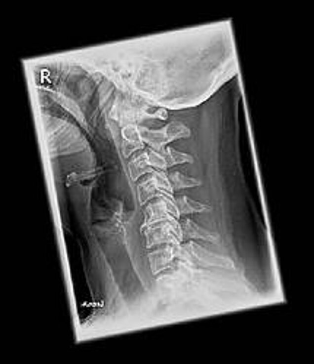
X-RAY
X-rays are the oldest and most commonly used medical imaging technique. X-rays were discovered in 1895 and first used to image human tissue in 1896. X-rays use ionizing radiation to produce images of a person’s internal structure by sending beams through the body. These are absorbed at different levels depending on the density of the tissue.
X-ray radiation can generate three kinds of medical images; conventional X-ray imaging, angiography and fluoroscopy.
Conventional X-ray imaging generates an image of a localised part of the body, which will be analysed for anatomical abnormalities.
This kind of imaging usually evaluates:
- The skeletal systems
- The oral cavity (bone and teeth)
- Any ingested objects
- The lungs
- The breast (Mammography)
- The digestive system.
Angiography uses X-rays in combination with a contrast agent (chemical substances used to enhance specific structures in images) in order to visualise blood vessels, particularly the coronary arteries.
Fluoroscopy uses X-rays to visualise the internal structure in real-time, providing moving images of the interior of an object, such as hearts beating or throats in the process of swallowing.
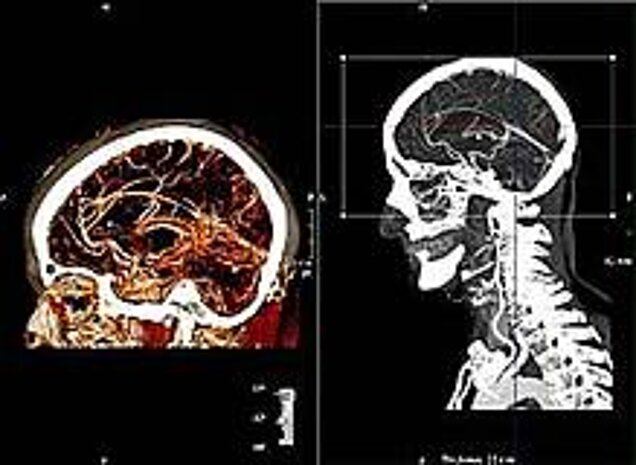
COMPUTED TOMOGRAPHY (CT)
Also commonly referred to as a CT scan, Computed Tomography is an imaging technique that combines multiple X-ray images taken from different angles. This produces detailed cross-sectional internal images. The first CT scanner for medical use dates from 1972.
The resulting images provide greater information than regular X-rays, allowing doctors to examine individual slices within the 3-D images. Contrast agents are commonly used in combination with CT scans to perform angiographies and other specific tissue examinations.
CT SCANS ARE OFTEN USED TO EVALUATE:
- Organs in the pelvis, chest and abdomen
- Colon health (CT colonography)
- Presence of tumours
- Pulmonary embolism (CT angiography)
- Abdominal aortic aneurysms (CT angiography)
- Spinal injuries
- Cardiology.
Technological improvements in CT such as dose modulation acquisition techniques and iterative reconstruction algorithms dramatically reduce the required X-Ray dose, improve hospital efficiency and clinical effectiveness and reduce costs.
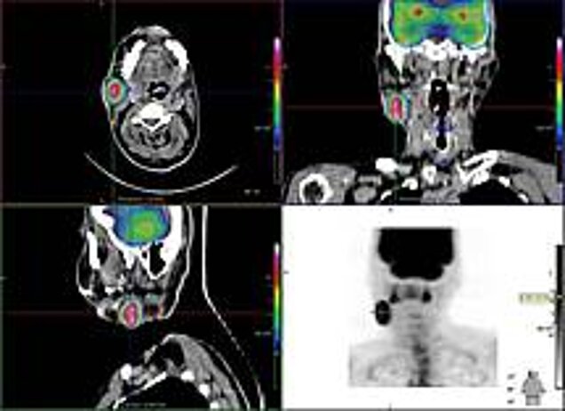
MOLECULAR IMAGING
Molecular Imaging is a diagnostic tool where metabolic processes can be visualised by administering small amounts of radioactive pharmaceuticals to patients. These accumulate in a specific part of the body in a controlled way.
Unlike other ionizing radiation techniques, which can only generate anatomical images, this technique generates functional images.
Some conditions initially have a physiological effect, rather than an anatomical change in the body. Molecular imaging allows for an earlier diagnosis.
Combining molecular imaging with CT or MRI images can provide clinicians with superior images. AIPES has developed a comprehensive tool on nuclear medicine. See website for further information.
Two specific technologies are used in nuclear medicine, namely POSITRON EMISSION TOMOGRAPHY (PET) and SINGLE PHOTON EMISSION TOMOGRAPHY (SPECT).
PET and SPECT are types of nuclear imagine techniques that provide physicians with information about how tissues and organs are functioning. In 1976 the first SPECT and PET scanners were developed going through 5 generations until what is in the market today.
These two techniques generate images that reveal the metabolism of the body, or the behaviour of certain particles, but cannot generate images of the anatomical structures. In order to provide a better image for the clinicians, molecular imaging can be combined with CT or MRI images so when the images are put together, the PET or SPECT images will be related to the anatomical structures obtained from the CT or MRI images.
PET and SPECT are often used as diagnostic and follow up images for:
- Neurological diseases such as Alzheimer’s and Multiple Sclerosis.
- Cancer.
- Heart disease.
NON IONIZING RADIATION
There are other medical imaging techniques that do not use ionizing radiation, using instead Magnetic fields, radio frequency or ultrasound radiation, that up to the moment have been proved to cause limited harm to the human body, therefore reducing the collateral damage to the surrounding tissue that is caused by ionizing radiation.
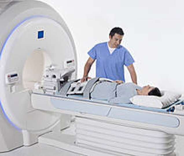
MAGNETIC RESONANCE IMAGING
Magnetic Resonance Imaging (MRI) is a technology that uses radio waves and a magnetic field to provide detailed images of organs and tissues. The first magnetic resonance image was taken in 1973 and the first MRI scanner for medical imaging was developed in 1977.
The type of radiation in this kind of imaging technique generates images of the soft tissues, omitting the bones. This characteristic has proven highly effective in diagnosing a number of conditions by showing the difference between normal and diseased tissues. MRI is often used to evaluate:
- Blood vessels
- Breasts
- Major organs
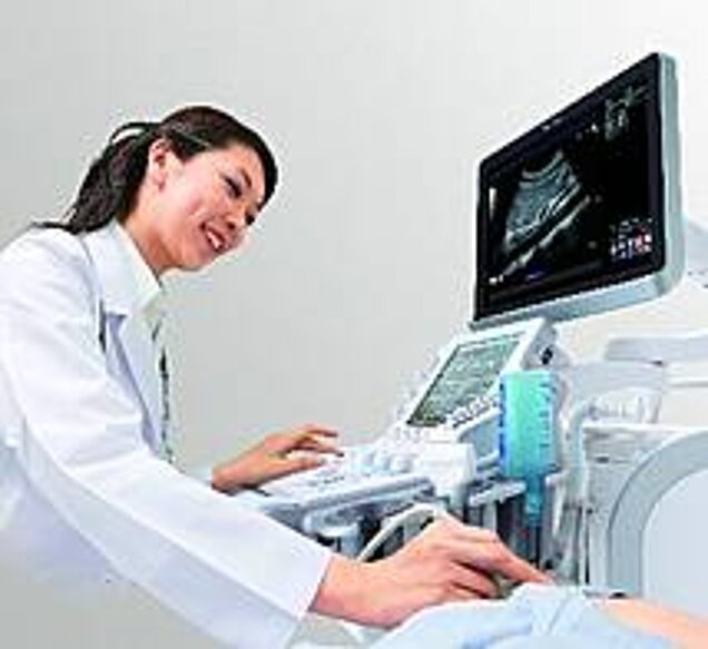
ULTRASOUND
Ultrasound have different applications, it can be used for therapy and muscle stimulation or as a diagnostic tool in medical imaging using an ultrasonographer. Diagnostic ultrasound, also known as medical sonography or ultrasonography, uses high frequency sound waves to create images of the inside of the body. The ultrasound machine sends sound waves into the body and is able to convert the returning sound, echoes, into a picture. The first image created with this technique was published in 1952. Ultrasound technology can also produce audible sounds of blood flow, allowing medical professionals to use both sounds and visuals to assess a patient’s health.
Ultrasound is often used to evaluate:
- Pregnancy
- Abnormalities in the heart and blood vessels
- Organs in the pelvis and abdomen
- Symptoms of pain, swelling and infection